
Nanonics, the leading provider of cutting-edge AFM technology, is proud to present its newest breakthrough in bio atomic force microscopy: the Hydra BioAFM(TM) featuring VISTA(TM) (Vivid Imaging Soft Touch AFM) mode. This innovation offers ultrasensitive single pN force mapping combined with the benefits of super-resolution optical/fluorescent imaging.
Live cell imaging with <100nm optical resolution and direct correlation with AFM topography is now possible. The finest structures, such as microvilli, can now be analyzed with on-line mechanics. The Hydra BioAFM, with its single or multi-probe capability, allows versatile optical integration with dual microscopes & side illumination. The system enables super-resolution methods like STED, PALM, STORM, and live cell NSOM with any available dye.
Key Features
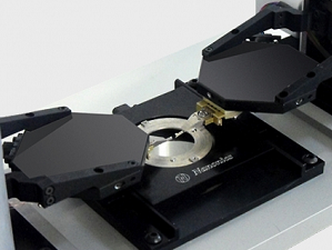
Optical Integration
 The Hydra BioAFM is designed for all forms of optical microscopy. The Hydra is the only BioAFM to integrate with either upright or inverted microscopes and dual (4pi) microscopes. Furthermore, side illumination for light sheet microscopy is available. The Hydra BioAFM has rendered obsolete solutions such as shuttling a sample from your AFM back and forth to an upright microscope. All of this is achieved with no compromise in force sensitivity and resolution.
(Seen above: Extended transparent glass probe with high Q Factor)
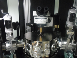
Correlated View
AFM overlaid with real-time CCD optical imaging Complete optical access combined with glass AFM probes provides for an unobstructed view Quick and easy probe-sample placement Quick selection of the area of interest for AFM imaging within the CCD image Generate beautiful 3D collages, correlating optical view with topographical and force information

Wide Versatility of Optical Configurations
- Upright
- Inverted
- Light sheet side illumination
- Combined 4 pi illumination (dual upright and inverted microscope)
- Brightfield & darkfield
- Phase contrast
- Epifluorescence
- TIRF
- Confocal fluorescence
- Raman and all forms of spectral imaging
- Non linear two, three photon
- SHG and STED imaging
See the Whole Picture
- Highly scattering tissue
- Bacterial cultures in Agar
- Opaque samples, such as microfluidic networks
Live Cell Imaging
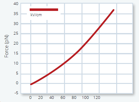 The Hydra BioAFM features VISTA(TM) (Vivid Imaging Soft Touch AFM) – enabling live cell imaging with the highest resolution. Combine highest resolution force mapping with controlled and known point of contact. VISTA offers: Live cell imaging with probes exhibiting Q factors in the thousands even in liquid Single pN force measurement – the highest force resolution of any commercial AFM Force spectroscopy – no Jump to Contact, allowing for investigating surface forces to the last nm from the membrane surface Ultralong extended probe tips with large aspect ratio and ideal angle for minimal cell perturbation 90μm Z range – the largest of any commercial AFM for imaging biological samples.
NSOM & Super-Resolution
NSOM - New Frontier in Live Cell Imaging
Near-field Scanning Optical Microscopy (NSOM) marks a paradigm shift in Super-resolution imaging. Live cell imaging with this powerful technique has been compromised in the past by stiff AFM cantilevers. Today Nanonics, with VISTA(TM), achieves the potential of live cell imaging with NSOM. Open New Frontiers of Super-resolution Optical Imaging (<100nm):
- Permits absorption and fluorescence
- Avoids the need for special dyes
- Correlates all optical contrast methods with 3D topography
- Allows highly confined zeptoliter illumination for fluorescence correlation spectroscopy (FCS) with physiologically relevant dye concentrations
- Enables AFM correlated FRET and dynamic FRAP with time resolutions previously unachievable
- Opens the door for the study of near-membrane phenomena with highly confined Z illumination
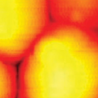
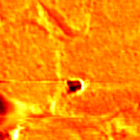
Seen below: An NSOM probe is seen approaching yeast cells
Top Left: AFM image of the Cells
Top Right: Transmission absorption NSOM
Bottom Left: Fluorescence NSOM
Bottom Right: NSOM probe seen from above with an upright microscope


Super-Resolution Optical Imaging
Over the last decade, a new powerful set of farfield optical tools has emerged for optical imaging of biological samples below the diffraction limit. These Super-resolution techniques have opened new horizons in the understanding of key biological phenomena. The Hydra BioAFM is compatible with all optical modes of Super-resolution. It permits the integration of a dual microscope for STED. PALM and STORM with total internal reflection prisms and an upright microscope are readily allowed. The Hydra BioAFM can combine AFM characterization with the following Super-resolution techniques:
- STED
- PALM
- STORM
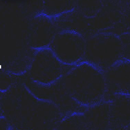
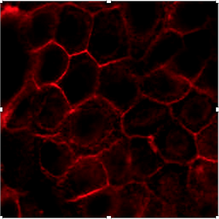
Seen below: Imaging of Living MDCK Cells With Dark Dots Indicating MicroVili Staining
Top Left: Super Resolution Absorption NSOM
Top Right: Linear Confocal
Bottom Left: Second Harmonic
Bottom Right: Two Photon

Super-Resolution Techniques Comparison

Specifications
AFM Specifications
1. Scanning Modes:Tip and sample scanning (Ability to scan either tip, sample or both)
2. Tip Scanning Range:
- MV 2000 BioAFM: XY=90 µm2 and Z=90 µm
- Hydra BioAFM: XY=40 µm2
3. Sample Scanning Range:
- MV 2000: XY=90 µm2 and Z=90 µm
- Hydra BioAFM: XY=90 µm2 and Z=90 µm
4. Closed Loop Scanner (optional): XY=100 µm2 and Z=20 µm
5. Feedback Modes:
- XY=100 µm2 and Z=20 µm
- High Q factor Tuning Fork feedback
- Optical feedback (optional)
Optical Modes of Operation
- NSOM/SNOM
- NSOM Fluorescence
- Confocal microscopes
- Raman Confocal microscopes
- Ability to integrate a variety of Super-resolution methods such as PALM/STORM/STED
Supported Modes
All standard SPM operating modes including:
- Force-distance spectroscopy
- SECM, SICM
- Nanolithography & Nanowriting
- MFM, EFM, Kelvin, Thermal, Electrical resistance, I-V curves etc.
Optical Integration
Optical Access: Clear optical access from above, below and side
Optical Microscope Configurations - Integration with any research grade optical microscope:
- Upright
- Inverted
- Dual optical microscopes (combined upright and inverted)
- Compatible with high NA optical objectives including water immersion objectives from both above and below
Accessories
- Liquid cell with seamless optical integration from above and below
- Optional PEEK liquid cell for caustic solutions
- Compatible with SECM, SICM
- Heating/cooling module using a programmable controller with mKelvin resolution
- Chamber for controlled environment with full optical integration for both upright and inverted microscopes
- Inlets for gas and humidity control
- Micro fluid control
Software
- Dedicated system control and data acquisition
- Customized scripts and synchronization of data by triggers in real time of imaging
- Hybrid optical/AFM mode for control and synchronization of hybrid imaging techniques
- User friendly software with video tutorials and on-line support by our experts
- Data analysis and processing software
Contact a Nanonics Specialist to Discuss Your Specific Needs
We are happy to answer all questions and inquiries
Images
Error


Videos
Live Cell Imaging
AFM Imaging of Neuroblastoma Cells
Nanolithography SPM Writing of a Protein
Deposition of Protein Nanoarray
Liposome Football
AFM Controlled Deposition of Cells and Subsequent AFM Imaging
Four Independently Controlled Probes
Environmental Control Chambers for Scanned Probe Microscopy
Configurations
Which Nanonics system configuration is right for your NSOM research?
Single Probe
- Compact design
- Versatile optical configurations
- XYZ range: 180μm
- Largest Z range of any commercial AFM
MultiProbe
- Multiple AFM probes for combined SPM measurements
- Compatible with standard biological μManipulators
- Keep your patch clamping pipette in place while imaging with AFM
Low Temperature
Publications
| 2018 | Association between biomechanical alterations and migratory ability of semaphorin-3A-treated thymocytes. | |
| Lins, M. P., da Silva, E. C. O., da Silva, G. R., de Souza, S. T., de Medeiros, N. C., da Silva Fonseca, E. J., & Smaniotto, S | Read the Abstract | |
| Biochimica et Biophysica Acta (BBA)-General Subjects. |
| 2017 | Nanosecond pulsed electric field induced changes in cell surface charge density. | |
| Dutta, D., Palmer, X.-L., Asmar, A., Stacey, M., & Qian, S. | Read the Abstract | |
| Micron, 100(April), 45–49. |
| 2017 | Characterization of CNL like protein fragment (CNL-LPF) from mature Lageneria siceraria seeds. | |
| Kumari, N., Chaturvedi, S. K., Khan, R., Sharma, A., Khan, R. H., & Yadav, S. | Read the Abstract | |
| International Journal of Biological Macromolecules. |
| 2017 | Adverse effect of CdTe quantum dots on the cell membrane of Bacillus subtilis: Insight from microscopy. | |
| Kumari, A., Khare, S. K., & Kundu, J. | Read the Abstract | |
| Nano-Structures & Nano-Objects 12 (2017): 19-26. |