
The MultiView 2000 series is an advanced single probe scanning probe microscope enabling a variety of modes of AFM/SPM/NSOM imaging. Nanonics has designed The MultiView 2000 for excellence in scanning probe microscopy while allowing for near-field and far-field optical NSOM/AFM Raman/TERS imaging without perturbation. The Multiview 2000 is the only commercially available instrument that offers both tip and sample scanning. This versatility is important for different operation modes where the user can now choose whether the sample or tip is static. The Multiview 2000 further offers the most stable feedback mechanism available in the form of normal force feedback with tuning fork actuation. This feedback mechanism offers the most stability, as well as laser-free operation for the most sensitive experiments.

Key Features
Designed for Ease of Use and Flexability
 Dual sample and tip scanning stages in the same head for ultimate flexibility is coupled with an optically friendly Nanonics 3D Flat ScanTM stage providing up to 85μ in X, Y and Z or 170μ in X, Y and Z using combined scanners. This scanner is also suitable for large samples with unconventional geometries. Closed loop is an option for positioning accuracy of 20nm. Additionally, a revolutionary, alignment-free feedback system provides the most sensitive tip-sample stability. Normal force feedback with tuning fork actuation provides no alignment, laser-free operation for improved optical signal to noise ratio, and ultra-sensitive force spectroscopy perfect for force and adhesion measurements. See here for more information and details about tuning fork feedback.
Compatible with a Wide Variety of Probes
 Nanonics provides a wide variety of probes that are compatible with the MultiView 2000TM, including: transparent, cantilevered probes, thermal probes, electrical probes (electrochemical probes, coax probes, glass insulated wire probes) and all third party silicon probes.
Largest Commercially Available Z-Range
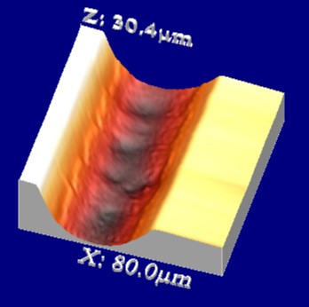
The large, 85μ X, Y and Z-range of the Nanonics 3D FlatscanTM makes it ideal for optical sectioning in confocal imaging. Used in this way, the MultiView 2000 integrates conventional far-field imaging, confocal microscopy, AFM, and near-field optics in a single system.
Any Optical Configuration
 The unique geometry of the MultiView 2000TM head and cantilever probes leaves the optical axis free both above and below the sample for integration with upright, inverted, and dual optical microscope configurations.
Irregular Sample Size Compatibility

Irregular sample sizes, whether odd in shape or large in size, can be easily placed on the sample stage.
 The sample can even be suspended from the unobstructed flat scanning sample stage in order to examine the edges of the sample.
NSOM Integrations
All Modes of NSOM
True Collection Mode
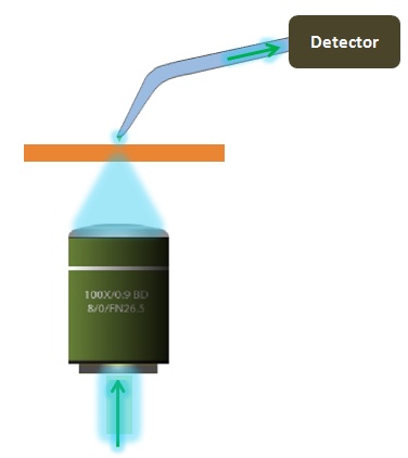 The MultiView 2000 offers both tip scanning and a sample scanning stage. A sample scanning stage enables easy and rapid alignment of the sample relative to the illumination source and ensures that the microscope optics are independent of the AFM scanner. A tip scanning stage means that the tip can be scanned while the illumination point (either from above or below) is held static. For an explanation of all modes of NSOM, please see here. This instrument design is ideal for true collection mode NSOM, where the light illuminates the sample, and then collected through the NSOM probe by a detector (see schematic on right).
True Reflection Mode
 In true reflection mode, light is introduced via the NSOM probe, and then collected by a detector above the probe as shown in the schematic below. The design of the MultiView series take advantage of transparent probes and separates the excitation and collection paths so that they don't affect each other. Other designs that take advantage of straight probes or apertured Si probes are significantly more challenging for reflection mode NSOM experiments. -More on different modes of NSOM can be found here
Apertureless and Scattering NSOM
Click below to learn more about the SpectraView 2500, ideal for apertureless and scattering NSOM
Completely flexible optical access - designed for NSOM use
This enables total flexibility in your NSOM setup, as the MultiView 2000 can be integrated with any optical microscope. Additionally, total optical access to sample and probe position from above is possible since the cantilever probe/scanner assembly does not obscure access
Transparent & cantilevered probes for the best NSOM performance
Nanonics is the global pioneer in glass probe manufacturing and has developed a whole suite of probes for all your NSOM applications featuring:
-Glass cantilevered normal force probes. These types of probes operate as standard robust AFM probes for easy, high-quality imaging of all samples. Additionally, the optical transparency of these probes provides unobstructed optical view from above and below
-Efficient Apertureless NSOM probes
-Fiber probes with metallic nanoparticles in tip
-Straight probes also available for shear force feedback measurements
See here for more information about Nanonics probes.
Raman Integrations
The Nanonics MV 2000 can be transparently integrated with ANY Raman System.
Free optical axis from top and bottom of upright and inverted optical microscopes for online AFM/Raman and TERS measurements
1. Direct Integration on the Stage of any Raman System

Nanonics MV 2000 - Renishaw Raman

Nanonics MV 2000 - Horiba XploRA Raman

Nanonics MV 2000 - Thermo Fisher Raman

Nanonics MV 2000 - Bruker Raman
2. Integration with an Additional Microscope


3. Integration with your Choice of Monochromater & CCD

4. Fiber Integration

SEM/FIB Integrations
Vacuum compatible SPM head for integration with SEM/FIB/ion beam systems
Clear electron and optical axes for on-line AFM/NSOM with SEM/FIB/ion beam operation

Nanonics MV 2000 - SEM FIB Integration

Exemplary Applications

Complete transparency of MultiView 2000 inside SEM chamber.
Environmental/Vaccum/Glove Box Chamber Integrations
- Compatible with high vacuum chambers with integration into optical microscopes and free optical axis from top and bottom
- Monitored humidity-control capabilities ranging from 5% - 95%
- Cooling to 4C and heating to 40C inside the chamber
- Inlets for additional environmental-control substances, including gas inlets
- Optical fiber inlets
- Sealed container designed to allows manipulation of the MultiView 2000 head in a Glove Box for controlled atmosphere of inert gas, vacuum, humidity and chemicals.

Environmental chamber suitable for Glove Box integration with free optical axis

Environmental chamber scheme side and top view

Flexible integration inside high vacuum chambers with free Z optical axis.
Liquid Cell Integrations
A new frontier in integrated microscopy has arrived with the introduction of a unique liquid cell AFM/SPM LC 2000 TM for the Nanonics AFM/SPM 2000 Confocal Microscope TM that is shown below. This microscope system with liquid cell in place allows for the first time a high power low working distance (3.5 mm) water immersion objective from above and oil immersion objective from below and permits temperature control. This allows for efficient collection of fluorescent and other light fully correlated with atomic force microscopy imaging.

Contact a Nanonics Specialist to Discuss Your Specific Needs
We are happy to answer all questions and inquiries
Images

6x6µm AFM image of a state-of-the art transistor

On-line correlating Raman map of the strained silicon 400cm-1 peak

AFM image of near-field cathodoluminescence from a GaN nanowire under ion beam excitation obtained with MultiView 2000 inside a SEM chamber.

On-line NSOM image obtained in collection mode, showing an evanescent light decay along the nanowire and light distribution at the nanowire output
Videos
Reflection NSOM
TERS
AFM Probe Approaching a Surface in a SEM
AFM Probe Scanning a Grating in a SEM
The Importance of Glass Probes
Optically Transparent SPM Scanner
Publications
Click here to see 100s of publications