NSOM (Near-field Scanning Optical Microscopy), sometimes referred to as SNOM, is the premier tool for imaging and manipulation of light on the nanoscale. It is the only super resoltuion optical method that captures the near-field of light distribution, allows for all forms of optical imaging, correlates light distirbution with topography, and is effective in all environments.
Nanonics offers instrumentation for unlimited NSOM, thereby enabling a broad set of applications for NSOM, both apertured and apertureless near-field imaging. In addition, Nanonics has pioneered multiprobe NSOM platforms that are effective for pump/probe optical transport phenomena on the nanoscale. Open new frontiers with this paradigm shift in super-resolution imaging in the 50-100nm range that allows investigation of both surface and near-surface phenomena.
Key Features
Nanoscale Near-field Optical Imaging
Nanoscale optical super-resolution of surfaces and near-surfaces, without out-of-focus light, correlated with topography and phase in all modes of imaging: absorption, reflection, collection, transmission, fluorescence, and other spectroscopies - in all environments!

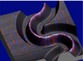
Collage of NSOM light distribution (colors) with topography of Ebessen plasmonic structure (Collection Mode)

Wire laser light distribution at two wavelength 805nm and 805.8nm
All Forms of Optical Imaging
Allows for all forms of optical imaging: absorption, reflection, collection and illumination with a point source as small as 50nm.

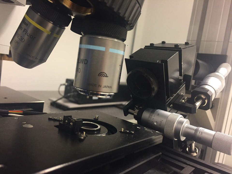
- Cantilevered glass AFM probes - exclusive for Nanonics systems
- Above and side views
- Compatibility with all third party probes

- High N/A with 1 mm working distance
Nanoillumination
Nanoillumination of a plasmonic waveguide—localized illumination without background


Correlation of Topography with Light Distribution
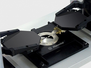
The simultaneous and independent SPM probe operation of up to 4 probes enables full characterization of all dimensions of samples and devices. All forms of SPM are enabled for each individual probe accompanied by total optical integration resulting in access to all modes including AFM, KPM, EFM, ANSOM, NSOM, STM, AFM-Raman, and TERS. Because these probes are designed for multiple probe operation, they can be brought to a nanometric separation distance thus ensuring that the same area is scanned with all the probes.
Imaging Below the Surface
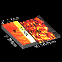
Reflection NSOM can see below surfaces in this state of the art Silicon chip of FITS structures

Topography

NSOM is one of the few near-surface techniques
Different Environments
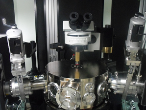
NSOM can be applied in all environments: from ambient conditions to cryogenic temperatures, and in liquid.

Probe completely immersed in the physiological solution while viewing from above with a water immersion objective

Microvilli in live MDCK cells with a cantilevered glass probe attached to tuning fork
Q factor in liquid 5000 normally 4 or 5 with beam bounce methods silicon cantilevers
Only in 2016 were very specialized silicon cantilevers able to image this structure and even these cantilevers could not approximate the ideal of glass probes and so were not able to measure the elasticity.
Modes

Speak with an application scientist
about how Apertureless NSOM
can advance your research

Speak with an application scientist
about how Reflection NSOM
can advance your research

Speak with an application scientist
about how Collection NSOM
can advance your research

Speak with an application scientist
about how Transmission NSOM
can advance your research

Speak with an application scientist
about how Fluorescence NSOM
can advance your research

Speak with an application scientist
about how Nano-Illumination NSOM
can advance your research
Images
Error


Contact a Nanonics Specialist to Discuss Your Specific Needs
We are happy to answer all questions and inquiries
Videos
Watch this video below and note the reflection of the point light source as it flickers. This is due to alterations in the near-field optical interaction, caused by scanning a semiconductor nanoelectronic chip. With their exposed tips, Nanonics cantilevered near-field apertured probes readily allow for a free optical axis for maximum collection of the reflected light signal above the probe, by a lens in a standard upright optical microscope.
Diffractive Optics and Nanodimensions
Reflection NSOM Near-Fileld Induced Flickering Tip Intensity
Two Probe NSOM
Close Approach of Two NSOM Tips
Four Independently Controlled Probes
Reflection NSOM Tip Intensity Modulation
Publications
2018
Photonic candle–focusing light using nano-bore optical fibers.
Schneidewind, H., Zeisberger, M., Plidschun, M., Weidlich, S., & Schmidt, M. A.
Optics Express, 26(24), 31706-31716. (2018).
2018
Near-field digital holography: a tool for plasmon phase imaging
Dvořák, P., Kvapil, M., Bouchal, P., Édes, Z., Šamořil, T., Hrtoň, M., ... & Sikola, T.
Nanoscale.
2018
Experimental study of metasurface-based nanoantennas array fabricated using heavy ion tracking for biochemistry sensing.
Cui, S., You, Y., Zhao, K., Fu, Y., Liu, J., Duan, J., & Zhu, S.
Sensors and Actuators B: Chemical.
2018
Observation of surface plasmon polaritons in 2D electron gas of surface electron accumulation in InN nanostructures
Madapu, K. K., Sivadasan, A. K., Baral, M., & Dhara, S.
Nanotechnology
2018
Integrated amorphous silicon-aluminum long-range surface plasmon polariton (LR-SPP) waveguides.
Sturlesi, B., Grajower, M., Mazurski, N., & Levy, U.
APL Photonics, 3(3), 036103.