
The Limits of Far Field Imaging
In the early 1870s, Ernst Abbe formulated a rigorous criterion for being able to resolve two objects in a light microscope: d > λ / (2sinθ) where d = the distance between the two objects, λ = the wavelength of the incident light, and 2θ = the angle through which the light is collected.
According to this equation, the best resolution achievable with optical light is about 200 nm.
With the introduction of NSOM (near-field scanning optical microscopy, also known as SNOM, scanning near-field optical microscopy), this limitation no longer exists, and optical resolution of < 50 nm can be achieved.

The Basic Principle of NSOM


Click above to enlarge
Light passes through a sub-wavelength diameter aperture and illuminates a sample that is placed within its near field, at a distance much less than the wavelength of the light. The resolution achieved is far better than that which conventional optical microscopes can attain.
A Brief History of NSOM
1928/1932
E.H. Synge proposes the idea of using a small aperture to image a surface with sub-wavelength resolution using optical light. For the small opening, he suggests using either a pinhole in a metal plate or a quartz cone that is coated with a metal except for at the tip. He discusses his theories with A. Einstein, who helps him develop his ideas. [E.H. Synge, "A suggested method for extending the microscopic resolution into the ultramicroscopic region" Phil. Mag. 6, 356 (1928); E.H. Synge, "An application of piezoelectricity to microscopy", Phil. Mag., 13, 297 (1932)].
1956
J.A. O'Keefe, a mathematician, proposes the concept of Near-Field Microscopy without knowing about Synge's earlier papers. However, he recognizes the practical difficulties of near field microscopy and writes the following about his proposal: "The realization of this proposal is rather remote, because of the difficulty providing for relative motion between the pinhole and the object, when the object must be brought so close to the pinhole." [J.A. O'Keefe, "Resolving power of visible light", J. of the Opt. Soc. of America, 46, 359 (1956)].
In the same year, Baez performs an experiment that acoustically demonstrates the principle of near field imaging. At a frequency of 2.4 kHz (כ = 14 cm), he shows that an object (his finger) smaller than the wavelength of the sound can be resolved.
1972
E.A. Ash and G. Nichols demonstrate כ / 60 resolution in a scanning near field microwave microscope using 3 cm radiation. [E.A. Ash and G. Nichols, "Super-resolution aperture scanning microscope", Nature 237, 510 (1972)].
1984
The first papers on the application of NSOM/SNOM appear. These papers are the first to show that NSOM/SNOM is a practical possibiltity, spurring the growth of this new scientific field. [A. Lewis, M. Isaacson, A. Harootunian and A. Murray, Ultramicroscopy 13, 227 (1984); D.W. Pohl, W. Denk and M. Lanz, APL 44, 651 (1984)].
The Basic Setup of NSOM
In order to make an NSOM/SNOM experiment, a point light source (1) must be brought near the surface that will be imaged (within nanometers). The point light source must then be scanned over the surface, without touching it (2), and the optical signal from the surface must be collected and detected (3).
1. There are a few different ways to obtain a point light source:
- One can use pulled or etched optical fibers (tapered optical fibers) that are coated with a metal except for at an aperture at the fiber's tip. The light is coupled into the fiber and is then emitted at the sub-wavelength (50 nm or larger) aperture of the fiber.
- Or, one can use a standard AFM cantilever with a hole in the center of the pyramidal tip. A laser is focused onto this hole, which is of sub-wavelength dimensions.
- Finally, the tip of a tapered pipette can be filled with a light emitting compound, which can then be excited either by light or by applying a voltage. It is also possible to use chemical luminescence.
The resolution of an NSOM/SNOM measurement is defined by the size of the point light source used (typically 50-100 nm).
2. The distance between the point light source and the sample surface is usually controlled through a feedback mechanism that is unrelated to the NSOM/SNOM signal. Currently, most instruments use one of the following two types of feedback:
- Normal force feedback (the standard feedback mode used in AFM), which enables one to perform experiments in contact and in intermittent contact mode. This feedback mechanism is only possible with cantilevered, tapered optical fibers and with AFM cantilevers with holes.
- Shear force feedback, or tuning fork feedback. The straight tip is mounted to a tuning fork, which is then oscillated at its resonance frequency. The amplitude of this oscillation is strongly dependent on the tip-surface distance, and it can be effectively used as a feedback signal.
Shear force imaging is not understood very well, and there are a lot of artifacts in the topographical images that one obtains using the method.
3. There are four possible modes of operation with NSOM/SNOM:
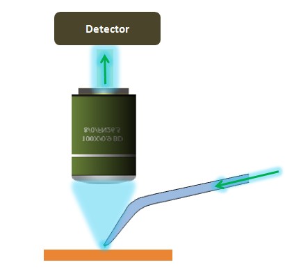
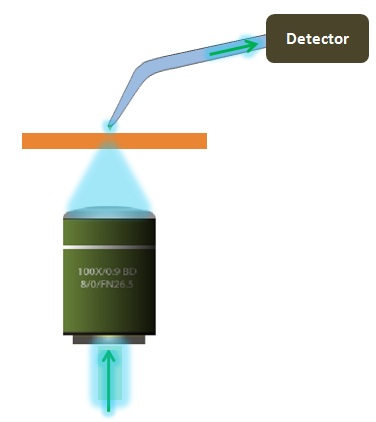
- Transmission mode imaging. The sample is illuminated through the probe, and the light passing through the sample is collected and detected.
- Reflection mode imaging. The sample is illuminated through the probe, and the light reflected from the sample surface is collected and detected.
- Collection mode imaging. The sample is illuminated with a macroscopic light source from the top or bottom, and the probe is used to collect the light from the sample surface.
- Illumination/collection mode imaging. The probe is used for both the illumination of the sample and for the collection of the reflected signal.

Detecting the collected light can be achieved with a wide variety of instruments: an Avalanche Photo Diode (APD), a Photomultiplier Tube (PMT), a CCD, or a spectrometer. The signals obtained by these detectors is then used to create an NSOM/SNOM image of the surface.
On the right, a schematic view of a transmission, normal force mode NSOM/SNOM setup with all the components required for its operation.

NSOM Modes


Advanced Methods
Professor Aaron Lewis, who was awarded the Rank Prize in optoelectronics for his pioneering development in near-field optics, reviews the basis and the history of near-field optics. He explains near-field optics within the context of such other super-resolution techniques as STED (Stimulated Emission Depletion Microscopy), PALM (Photoactivated Localization Microscopy), STORM (Stochastic Optical Reconstruction Microscopy) and SIM (Structured Illumination Microscopy). The lecture shows how important excellence in AFM (Atomic Force Microscopy) is to the correlation of the optical distribution with critical topographic features such as the gradient of morphological changes and other such structurally based AFM information. Furthermore, the lecture clearly shows the importance of transparent integration of NSOM, AFM and Optical Lens Based imaging which is only possible due to an integrated, flexible and open design philosophy that allows for an optically non-obscuring geometry of the probe and a non-obscuring structure of the AFM instrument.
Contact a Nanonics Specialist to Discuss Your Specific Needs
We are happy to answer all questions and inquiries