

Super User
AFM Raman of Si SiO2

 |
 |
Online AFM/Raman images of an Si/SiO2 grid shows a high lateral resolution on the Raman map. Shown on the left is the topographic image; on the right is the online Raman map of the 520cm-1 band’s intensity.
-
Nanonics’ MultiViewTM SPM systems – with their 3D FlatScanTM scanners and cantilevered glass probes – provide a free optical axis for a friendly online combination of AFM and Raman spectroscopy using true confocal optical microscopes, including upright microscopes.
-
Online AFM/Raman allows for direct and true correlation between structural and chemical information of the inspected samples. In addition, it improves the Raman’s lateral resolution by removing out-of-focus light through the accurate maintenance of sample-objective distance using the AFM tip. Finally, online AFM/Raman corrects tilts caused by the normal tilting of samples.
-
Glass probes are critical for Si-based samples, to prevent any background caused by the tip, as in most standard AFM systems that use Si cantilevered probes.
-
A Tip & Sample-Scanning AFM system is ideal for this type of application, to obtain tip/laser positioning for accurate AFM/Raman correlation. Complete correlation is obtained, without the miss-matching that can occur due to the switching of microscope objectives.
Ideal Systems for this Application:


AFM Raman of Name Card

Simultaneously Obtained Topography and Raman Images of Name Card
 |
 |
 |
|
Topography image |
Raman image at 1547 cm-1 |
Raman image at 954 cm-1 |
AFM and NSOM of A Multi Mode Fibre


A) AFM topographic image of a cleaved multimode optical fiber obtained with AFM/NSOM probe.
B) A correlating NSOM image in collection mode obtained simultaneously with (A).
C) A 3D collage AFM/NSOM of the output optical distribution with exact correspondence to the surface’s topography.
-
Near-field optical distribution of a multimode optical fiber launched with 532nm laser.
-
The Nanonics' 3D FlatScanTM stage allows for vertical mounting of the optical
fiber with a flexible geometry for optical microscopy integrations. -
The sample is kept stationary along the scan to prevent any disturbance of the light propagation through the fiber. Nanonics systems with Tip-Scanning capabilities are ideal in such applications for true profiling of the optical output.
-
The AFM/NSOM fully correlated imaging is based on tuning fork feedback in normal mode.
Nanonics Nano3D Distortion Free, Near-field/Far-field Beam
Profiler
This novel product provides high precision, 50nm optical resolution with simultaneous topography and without deconvolution.
-
Unprecedented error-free profiling of divergent sources with no detector saturation or beam attenuation.
-
No non-uniformities or astigmatisms in profiling active or passive sources such as VCSELs, AWGs, Ultrasmall Mode Field Diameter Lensed Fibers etc.
-
Seamless complex beam structures 3D profiles from the near-field to the far-field with overlapping fields of view.
Ideal systems for this application:
 |
An optical fiber vertically mounted on MultiView 2000TM SPM head integrated with an upright optical microscope |
TFT in Liquid Crystal Image

Thin Film Transistor in Liquid Crystal Display
 |
 |
|
| 50 x 50 micron AFM Topgraphy | Simultaneously produced NSOM image | |
| |
||
 |
|
AFM, NSOM and Capicatance SRAM Image

SRAM
AFM, NSOM and Capacitance Imaging
The first simultaneous NSOM/Capacitance ever to be produced.
 |
 |
|||||||
| 10x10 micron AFM Topgraphy | NSOM Image of the same region | |||||||
|

|

High Resolution NSOM Imaging in Reflection mode

A) Topographic AFM image of a Photonics Band Gap (PBG) structure, imaged with a 150nm AFM/NSOM cantilevered probe.
B) Online correlated NSOM image of the PBG structure at (A). Imaged with the same probe in reflection mode at a 532nm laser wavelength.
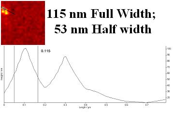
C) NSOM line profile shows a lateral resolution of 100nm in reflection mode.
Ideal systems for this application:
1. MultiView 1000
|


PEO Spherulite Imaging

|


High Resolution NSOM Imaging of Au Grid

|


Murine Stem Cell Imageing

|
||||||||||||||||||||||||||


