Super User

Super User

Surface plasmons excitation with STM probe and collection with apertured & apertureless NSOM photon tunneling probes

MV4000 Dual Probe function provides a flexible platform for simultanous STM and NSOM functionality. Such platform is ideal for surface plasmons (SP) excitation with an STM probe and effective imaging of the SP both in aperture NSOM and apertureless via photon tunneling process. 
The following scheme describes the dual probe setup for excitation of surface plasmons on 35nm gold coated surface. The second probe is an NSOM probe use for localized AFM/NSOM imaging of the generated surface plasmons at nanometric close proximity from the STM excitation probe. 
 

Left: Dual probe Setup for SPP excitation with STM probe and near-field imaging with a second NSOM probe. The multiprobe setup allows for clear optical access from above and below without any obscuration. Right: An optical microscope image from above shows dual probe STM/NSOM in during operation. Cantilevered bent probes allows for clear optical access and for multiprobe operation at close proximity. 

AFM NSOM Imaging of Surface Plasmons excited with STM Probe 

Simultaneous AFM-NSOM imaging of surface plasmons with an apertured NSOM probe. The cantilever NSOM and STM probes geometry enables bringing the NSOM probe to a close proximity from the excitation STM probe and thus allows mapping of the SP the poitn where they are generated.  In addition, the MV 4000 system incorporates sample scanning and tip scanning piezo stages and thus allows for flexible an accurate positioning of both probes until achieving "soft contact" between the probes. Such a procedure of two probe distance adjustment is performed while the two probes arekep in feedback with the sample and thus allows for safe probe manipulation

The image below shows the results of the above protocols obtained by the NSOM probes in probe scanning mode.


Simultanous AFM Height (Left) and NSOM (Right) images performed with the aperture NSOM probe starting from the STM tip position (placed at a stationary position at the bottom side of the above image)


Ultra Sensitive Tuning Fork for AFM/STM Feedback

MV4000 multiprobe system uses ultrasensitive Tuning Fork feedback which is ideal for such combined AFM and STM feedback. The tuning fork high force sensitivity and lack of jump-to-contact allows for defined tip-sample interaction down to close proximity from the sample (<1nm) without any force discontinuity (which does occur in optical feedback AFM techniques, during to jump-to-contact and ringing adhesion effects). Such distance stability permits obtaining simultaneous AFM and Tunneling

Ultrasensitive tuning fork AFM-STM feedback with Normal Force AFM/NSOM cantilevered and exposed probes.


Unique AFM-STM probes

Nanonics uniquely provides probes which are able to scan with either AFM or STM feedback using the same probe with easy switching between modes

AFM STM Probe acting in Normal Force with tuning fork feedback


Single and multiple atomic steps imaging with AFM and STM feedback. Line profiles of HOPG steps with AFM feedback (middle) and STM feedback (Right) performed with the same probe.

Nanonics MultiView AFM/STM system has been used for obtaining Work Function information on Q-Dot using Tunneling Feedback.  

Published:  Nano Lett. 2013, 13, 2338−2345

Click here for more information on the Nanonics MultiView 4000 system


One of the most powerful applications of multiprobe instrumentation for near field optical measurements is the novel capability for pump/probe experiments.  In NSOM pump/probe experiments, an optically active system is excited in the near-field (or pumped), and then mapped (or probed/detected) in the optical near fieldsimultaneously.  These optical pump-probe measurements enable the measurement of optical properties of nanostructures with the most accuracy and highest resolution.

However, now that there are two near field optical signals to measure, the complexity of interpreting these dual near-field type measurements is higher. Authors Angela Klein et al. of Institute of Applied Physics in Jena [Nano Letters, 2014]shed light specifically on exploring the polarization characteristics of the excited and detected light in dual-probe NSOM experiments using a Multiview 4000 system.  They find that the cantilever fiber probes, which are the conventional geometries of probes used in NSOM measurements, can both emit polarized light and function as polarization sensitive detectors.  Specifically, they make measurements on surface plasmon polaritons and find dipole-like SPP emission from the tips.

They conduct direct near field measurements  of dipole-like Surface Plasmon Polaritons (SPP) emission from a cantilevered aperture NSOM probe.  They study the polarization characteristics of Nanonics cantilevered single mode (SM) and multimode (MM) aperture NSOM probes in far-field and near-field.

A cantilevered SM NSOM probe with a high polarization factor was used for excitation while a MM cantilevered NSOM probe was used for collection. The experiment was done on a gold coated substrate.

This paper demonstrates that a cantilevered NSOM probe not only acts as SPP dipole, but can also be used as a polarization sensitive near–field detector.

In Brief: The MultiView 4000 Nanonics MultiProbe NSOM is the only tool that can allow for such kinds of experiments.  This microscope also assesses and confirms the polarization characteristics of cantilevered NSOM probes which leads to a better understanding of NSOM measurements.

PublishedNanoLetters, 2014

Click here for more information on the Nanonics MultiView 4000 system


Tuesday, 03 February 2015 19:42

Dual probe NSOM on plasmonic metasurfaces

 Authors J.S. Clausen and colleagues, from the University of Denmark, report on a novel way to implement structural colors into plastic products for daily consumer use.  Structural colors offer some significant advantages over current pigment-based coloring by reducing the materials needed and new opportunities for recycling and sustainability.  The authors create structural colors here from metal disks on top of dielectric pillars that are hovering above a holey metal film composed of aluminum. They then use the dispersion of the surface plasmon polaritons (SPPs) supported by the metal-dielectric interface of the holey film to generate the colors; a schematic of the structure is shown below where the final protective coating is then added to minimize environmental contamination.  By using aluminum (instead of gold or silver), the SPPs supported by aluminum to improve the angle independence in the color observation, as well as other improvements.  The authors use a dual probe Multiview 4000 to characterize the SPP modes with excitation probe with a 100nm aperture diameter held fixed and a detection probe with a 200nm aperture diameter scanned away from the excitation.

Published:  Nano Letters 2014, 14, 4499-4504

Click here for more information on the Nanonics MultiView 4000 system


Tuesday, 03 February 2015 19:41

Multiprobe Nanoindentation

Novel Approach to Nanoindentation Using a Multiprobe System

Authors Eyup Cinar and Ferat Sahin  describe a unique approach to nanoindentation that utilizes a MultiView 4000 multiprobe setup with tuning fork technology to conduct AFM-based nanoindentation with power advantages for measurements on soft and compliant materials.  Their method relies on tuning fork actuation and a frequency-based feedback (instead of the classical amplitude based feedback), which enables a very precise measurement of tip-sample interactions that avoids instabilities such as adhesion ringing and jump to contact that plague conventional measurements.

In this novel and exciting method, a dual-probe setup (schematic shown below followed by actual setup) is implemented where one probe is a diamond-tipped indentation probe while the second AFM probe measures the displacement of the indentation probe.  The tuning fork actuation controls the motion of the diamond-tip probe, thereby bring unprecedented stability to this measurement.


The authors demonstrate initial results showing proof-of-concept of this approach to nanoindentation with indents on a silicon sample where the stage positions of the 2 probes is successfully monitored and force vs. displacement curves are collected as shown below.  They further simulate their results with finite element analysis that shows good agreement from the simulated level of force and experimentally obtained force for a given penetration depth.  These preliminary results demonstrate the exciting potential of this approach to nanoindentation, and further experiments are already underway to try this method on a variety of samples.  

Published:  IEEE Nano, Sahin and Cinar 2014

Click here for more information on the Nanonics MultiView 4000 system


Tuesday, 03 February 2015 19:39

NSOM on periodic ferroelectric domains


Collection mode and Reflection mode NSOM on ferroelectrics

Authors Camarillo and colleagues from Universidad of Autonoma de Madrid, Univ of Mexico, and Univ of Glasgow, study the behavior  of various periodic ferroelectric domains with NSOM to probe these periodically poled structures as optical superlattices.  They used a Multiview 2000 in both collective and reflection mode, finding a correlation in the NSOM signal intensity with ferroelectric domain structure.   Quasi-periodic ferroelectric domains structures act as optical super=lattices as a result of their refractive index modulation along the domain structure. The authors use the NSOM results to understand the refractive index modulation in their structures.

In the image below, AFM and NSOM in reflection mode were collected simultaneously using a CW laser beam from a Nd:YAG laser coupling into the NSOM probe, and detected by an avalanche photodiode (APD).  The domains that are topographically depressed (dark brown regions in (a)  ) showing a higher NSOM intensity than the domains that are topographically elevated.  Thus, they surmise that the topographically depressed domains are more effective as waveguides than the topographically elevated domains, suggesting that the refractive index value is higher in the topographically depressed regions.

In a ferroelectric structure fabricated in another way, the authors observe a different structure as seen in the AFM topography (a) and NSOM (b) image obtained here in collection mode.   Again, the areas of higher NSOM intensity are correlated to topographically depressed regions in the AFM image; though the topography in the AFM is not very strong, the authors surmise this is a result of polishing.  These observations are important to understanding the optical behavior of ferroelectric crystlas, which have potential application for nonlinear optics.


Published:  Ferroelectrics, 467 (p.6-12),  (2014)

Click here for more information on the MultiView 2000 system

Tuesday, 03 February 2015 19:38

Arbitrary bending of plasmonic light waves

Arbitrary bending plasmonic light waves

Plasmonic light waves along arbitrary curves

The ability to control and guide plasmonic light waves could present significant opportunities in photonics and electronics applications for nanoscale-on-chip technologies and subwavelength optical devices.  Different kinds of plasmonic beams have been observed including those that preserve their spatial shape with propagation (“nonspreading”) and those that propagate along curved trajectories (“self-accelerating”).  By conducting numerical simulations and direct experiments with NSOM using a Nanonics Multiview 2000 system, authors Itai Epstein and Ady Arie generate surface plasmon beams that propagate along arbitrary curved trajectories.  These beams have important applications such as enabling trapping/guiding of microparticles or bypassing a curve of concentrated light.


Published:  Physical Review Letters, 112, 023902 (2014)

Click here for more information on the Nanonics MultiView 2000 system

Tuesday, 03 February 2015 19:33

TERS detection of membrane receptors

The ability to obtain chemical information about various receptors within cell membranes could advance efforts for drug development that target disease associated with receptor activity.  A key class of cell membrane proteins associated with cell adhesion, differentiation, and growth are integrin receptrs. In this work, TERS conducted with a Nanonics Multiview 4000 selectively detects the Raman spectrum of the integrin receptor in the cellular membrane.

Authors Hao Wang and Zachary Schultz of University of Notre Dame [ChemPhysChem 2014]use nanoparticles functionalized with ligands to bind with intact cell membranes.  The nanoparticle then functions as an enhancing nanostructure enabling TERS measurements of the Raman spectra of the ligands.  The authors conduct TERS to detect the signal of the amino acid within the integrin receptor and thus elucidate the structure of the receptor’s binding site.  This demonstrates the powerful utility of TERS for obtaining sensitive chemical information about amino acids relevant to chemical processes within cellular membrane proteins.

TERS setup for imaging nanoparticle functionalized ligands

Published:  H. Wang et al., 2014 ChemPhysChem   

Click here for more information on the Nanonics MultiView 4000 system

Tuesday, 03 February 2015 19:33

AFM-Raman of nanoporous carbon from lignin

This paper by Spanish researchers Navarro-Suarez et al.  describes the development of a new technology to produce lignin-based nanoporous carbon with narrow and tunable pores. This novel material was prepared by chemical activation of natural lignin at 900˚C, and it was tested as an electrode material for electrical double-layer capacitors.

Characterization of the morphological, chemical and electrical properties of this new material was done by different methods including SEM, HRTEM, EDX, electrochemical analysis and Raman microscopy.  Raman characterization was performed with a Nanonics Multiview 2000 integrated with a Raman spectrometer. Such an integrated system enables characterization of morphological properties with simultaneous chemical characterization.  

The authors shows that there are ordered and disordered carbon regions and presence of few layer graphene (FLG) embedded into the amorphous carbon matrix.  The Raman spectral imaging and ratio between Iand IGRaman peaks of carbon confirmed SEM and HRTEM morphological results.  Further electrochemical studies showed that capacitance value of 6.87 Fg-1 can be obtained.

Thus, the obtained results demonstrated that nonporous carbon with tunable pore size can be achieved by using a natural material such as lignin and can be effectively used as electrodes material in energy storage applications. 

Distribution obtained by Raman spectral imaging of the ordered and disordered areas inside a particle for samples (a) 2, (b) 4 and (c) 6.

Color scale on the right varies with the ID/IG values from 0.0 (dark blue) to 1.2 (dark red).

Published:  RSC Advances, 2014, 4, 48336-48343

Click here for more information on the MultiView 2000 system


Near Field Scanning Microscopy has great potential in the study and characterization of 3D and 2D plasmonic metamaterials and metasurfaces.   In this paper, a Nanonics MultiView 4000 Dual probe Near-field Scanning Optical Microscope was used to investigate two phenomena in plasmonic metasurfaces (subwavelength antenna like pattern in ultrathin silver film) showing:

  1. Extraordinary suppressed transmission ( EOST) in metasurfaces

  2. Bright (radiative) and dark (non-radiative) plasmonic modes propagation in metasurfaces

The Nanonics MultiView 4000 Scanning Probe Microscope is ideally suited for complete characterization of the dark mode propagation.  One NSOM probe injects the light to excite the dark (non-radiative) modes, while the second NSOM probe scans in the vicinity of the illumination probe, and detects the dark modes distribution.

The NSOM results obtained demonstrate that far-field radiation resonantly excites antenna-like (bright) modes that are localized on the metal ridges. The re-radiation of these modes into far-field interferes destructively with the transmitted wave, thus almost completely suppressing transmission.  In contrast, a second type of mode, (dark) bound mode Surface Plasmon Polarities (SPPs) launched from the NSOM tip, propagate well across the metasurface, preferentially perpendicular to the grating lines.

Idea in Brief: It was shown that dark modes cannot be excited in the far-field, and instead NSOM tips are the best source for excitation and probing of the non-radiative modes, which do not interact with the far-field.  


Published:  Advanced Optical Materials, 2014, S. Dobmann et al.

Click here for more information on the MultiView 4000 system

Tuning forks for ultrasensitive force spectroscopy measurements

Link to PDF version of this app note

Introduction

Standard AFM-based methods to monitor mechanical properties of materials such as elasticity and adhesion are based on beam bounce or optical beam deflection technology (see Figure below).  In this technology a laser beam bounces off the reflective back end of an appropriately chosen cantilever that is sufficiently soft to probe the material of interest  The laser beam then tracks the bending of the cantilever or its amplitude as a surface is being approached , which is then analyzed to understand the mechanical properties of the material of interest.

With such an approach there are two major problems seen diagrammatically below.  This schematic shows the tip-sample force as a function of time with two major sources of instability:  the jump or snap into contact resulting in a discontinuity, which happens upon the approach, and then a second discontinuity at the point of withdrawal resulting in uncontrolled ringing of the cantilever.  This is called Adhesion Ringing and is due to the fact that the adhesion force between the tip and sample is much stronger than the soft cantilever.  Thus, the adhesion of the sample in effect plucks the soft cantilever like the string of a guitar, resulting in uncontrolled ringing that has to die down.


A better controlled experiment would include a smooth approach and retract by the tip so that the interaction is described by a smooth mathematical function as indicated by the dotted line.

An improved approach to force spectroscopy:  tuning fork actuation and frequency based feedback

One way to avoid these discontinuity problems in the tip-sample interactions is to use the cantilever’s oscillation frequency instead of its amplitude as the feedback parameter.  Very stiff cantilevers with very high Q oscillations are typically used in such experiments.  The sharp frequency spectrum is very sensitive to small changes in the frequency imposed by ultrasmall forces of a tip interacting with a surface.  This method is called Frequency Modulation. 

An ideal example of a stiff device that is even self-oscillating (since it is formed of a piezo material) is a tuning fork, which also has very sharp resonance The higher the device Q the higher its sensitivity to any external perturbation such as surface forces. Thus, Frequency Modulation with tuning forks is ideal for force spectroscopy. 

This method has been implemented among varied research groups1 1b, 2and have validated the fundamental force limits of a tuning fork that were estimated to be less than a pN nearly a decade ago3 .  A Nanonics instrument recently demonstrated force sensitivity as predicted by Grober et al. in a beautiful study by Kohlgraf-Owens et al.4

These efforts have now been complemented by the pioneering theory of Sader and Jarvis5 that has shown theoretically that it is possible to derive accurate formulas for the force versus frequency in such Frequency Modulation methods.

Thus, tuning forks whose force constants are many orders of magnitude larger than standard silicon cantilevers have significant advantages in force spectroscopy measurements over conventional cantilevers.  By using such stiff tuning forks, the problems in classical silicon cantilevers of jump to contact and adhesion ringing are avoided.  Tuning forks provide high sensitivity especially in the region of very close tip-sample contact.  This region could not be addressed previously because of the discontinuities in the measurement but is now accessible..  Tuning fork feedback also provides other advantages, such as in electrical measurements, where in many semiconductor devices the feedback laser causes induction of carriers.  Finally, tuning fork feedback does not employ a laser so that there is no laser feedback interference for electrical and optical spectroscopy measurements.

Deriving model parameters from tuning fork based force spectroscopy

Tuning forks have force constants of 2,400 N/m and so their Q is much higher  than the tens or hundreds of N/m force constants of rectangular cantilevers.  The DMT model can be used to model force curves collected with tuning forks.

Finteraction is the force between the tip and sample

E* is the reduced elastic modulus

R is the tip radius

d0 is the surface rest position

Fadh is  an adhesion force

Discontinuities and hysteresis in the force curve can affect accurate measurement of the parameters that go into the DMT model.  A jump to contact instability prevents direct measurement of “do” and so the Finteraction between the tip and the sample will also be complicated.  Furthermore, if there is adhesion ringing then similarly the adhesion force can be difficult to assess. 

Determining the DMT model parameters using a tuning-fork based setup is straightforward.  With frequency used as the feedback parameter, amplitude can now be monitored as a separate independent quantity.  When a tuning fork with a tip approaches the surface with frequency feedback, the oscillation amplitude is measured independently.  Thus the point at which the tip touches the surface is observed immediately since one sees an amplitude change independent of any effect of feedback.  Furthermore, this occurs without any jump to contact so one knows the exact point of contact or “do” in the equation above. 

This is shown practically in the Amplitude vs Distance graph below.  As the probe approaches the surface from the right on the graph, from I, the point at which the slope abruptly changes, T, is the point of contact or “do”.  The intersection of the straight line I T and the straight line TM now define “do”.  M is the point of greatest penetration or indentation.  Similarly the point of retraction from the surface without ringing is readily and smoothly determined by the intersections of MR with the straight line RF.


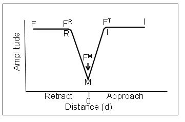
The forces measured at T (FT) M (FM) and R (FR) are the Finteraction, 

Fmaximum or peak force and Fadhfrom Sader and Jarvis [J. E. Sader and S. P. Jarvis.5  We now have accurate formulas for the changes in Q, which is related to frequency, during the force distance curves.  Therefore, the forces at T, M and R can readily be determined even in ultra close proximity to the surface without any jump to contact or adhesion ringing and all elements of the DMT equation are known experimentally.  Note that the amplitude distance curves do not have to be symmetric.  They are only symmetric when there is no adhesion force.    

Nanonics in collaboration with the Continuum Mechanics and Material Theory Faculty at the Technical Univeristy of Berlin has tested the implementation of the equations of Sader and Jarvis.  These tests are based on extensive experiments with calibrated micromechanical suspended cantilevers with accurately determined force constants both theoretically and by other experimental methods.  An example of such measurements is shown below. 


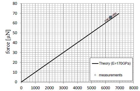
The microbeams allow for the imposition of relatively large forces that enable direct comparison of the beam bounce method with theoretical results and with the tuning fork measurements. The solid curve above is a theoretical curve of the Force.  The measurement points are shown and the data compare well with the theoretical results.     

Now let us compare the tuning fork results on the same cantilever on the graph below.

The blue curve is obtained with the tip at the point where the microbeam is attached to the bulk silicon support where any bending detected is the result of the probe and can be used as a correction if needed.  The red curve is at the free end of the microbeam and the force is calculated with the Nanonics Software by measuring the frequency change and applying the Sader and Jarvis equations.  The yellow line is then generated by subtracting the extrapolated blue curve from the red curve; the blue curve can only be obtained at low force without breaking the tip since this curve is obtained at the inflexible point of the connection of the microbeam to the silicon support.  This removes any effect of probe bending which is in any case very small in this case with such a large force constant.

Now let us compare the slope of the yellow curve in the tuning fork measurements with what was measured by the beam bounce.  They are identical.  Thus, the Sader and Jarvis equations as implemented by the Nanonics program accurately determine the known force constant of these test microbeams with the curves crossing zero as would be predicted for 0 Force. 

Thus, today Nanonics is pleased to allow our customers a level of accurate force measurements with a variety of functional probes (optical, electrical, thermal, chemical deposition etc) that has till today been impossible to achieve by other techniques.

References

1.            (a) Ternes, M.; Lutz, C. P.; Hirjibehedin, C. F.; Giessibl, F. J.; Heinrich, A. J., The force needed to move an atom on a surface. Science 2008, 319 (5866), 1066; (b) Gross, L.; Mohn, F.; Moll, N.; Liljeroth, P.; Meyer, G., The Chemical structure of a molecule resolves by atomic force microscopy. Science 2009, 325.

2.            Albers, B. J., Three-dimensional imaging of short-range chemical forces with picometer resolution. Nature Nanotechnology 2009, 4, 307.

3.            Grober, R. D., Fundamental limits to force detection using quartz tuning forks. Review of Scientific Instruments 2000, 71, 2776.

4.            Kohlgraf-Owens, D. C., Mapping the mechanical action of light. Phys Review A 2011, 84, 011807R.

5.            Sader, J. E.; Jarvis, S. P., Accurate formulas for interaction force and energy in frequency modulation force spectroscopy. Applied Physics Letters 2004, 84, 1801.